Podocytes Form Which Of The Following
Podocytes Form Which Of The Following - They are interconnected by slit diaphragms and cover the exterior basement membrane surface of the glomerular capillary. Web in general, the pupa is formed mainly from epithelial cells such as terminal cells and podocytes, which are highly specialized for ultrafiltration of body fluids. Novel molecular markers of the podocyte are identified. They form extensions (foot processes) that completely surround the glomerular capillaries and play a role in restricting the passage of plasma proteins into the urine. Web the podocyte's response to injury: In the healthy kidney, specialized cells called podocytes form a sophisticated blood filtration apparatus that allows excretion of wastes and excess fluid from the blood while preventing loss of proteins such as albumin. Single gene disorders affecting the slit diaphragm. Web the main biologic function of podocytes is to restrict the passage of albumin and other key proteins to the blood space within the glomerular capillaries and to prevent their passage into the extracapillary urinary space. When it comes to glomerular filtration, podocytes play an active role in preventing plasma proteins from entering the urinary ultrafiltrate by providing a barrier comprising filtration slits between. The injury and death of glomerular podocytes are the keys to dkd. The injury and death of glomerular podocytes are the keys to dkd. Recent studies point to an important role of podocytes in the physiology and pathophysiology of the glomerulus. Glomerular visceral epithelial cells, also termed podocytes, are highly specialized epithelial cells that cover the outer aspect of the glomerular basement membrane. The primary urine is produced from body fluids (interstitial. The injury and death of glomerular podocytes are the keys to dkd. Web podocytes are specialized, terminally differentiated epithelial cells in the kidney, located on the outermost layer of the glomerulus. Single gene disorders affecting the slit diaphragm. Mesangial cells help keep the basement membrane clean by engulfing macromolecules caught in its basement membrane. Web a) in one minute. Web structurally, podocytes form major processes and smaller foot processes. Podocytes have a crucial role in maintaining the glomerular filtration barrier and their loss leads to glomerular. They function as a critical size and charge barrier to prevent proteinuria. Web in general, the pupa is formed mainly from epithelial cells such as terminal cells and podocytes, which are highly specialized. Web the main biologic function of podocytes is to restrict the passage of albumin and other key proteins to the blood space within the glomerular capillaries and to prevent their passage into the extracapillary urinary space. In addition, the multifunctional features of this exceptional cell type are better characterized, identifying specific neuronal, phagocytic and muscle traits. These cells play an. Novel molecular markers of the podocyte are identified. Web podocytes are terminally differentiated cells of the kidney glomerulus that are essential for the integrity of the kidney filter. Pronunciation of podocyte with 2 audio pronunciations, 1 meaning, 4 translations and more for podocyte. Their function is primarily based on their intricate structure, which includes foot processes. Web podocytes are specialized. To operate effectively, this filter is under substantial hydrostatic mechanical pressure. Web podocytes are highly specialized cells of the kidney glomerulus that wrap around capillaries and that neighbor cells of the bowman's capsule. In addition, the multifunctional features of this exceptional cell type are better characterized, identifying specific neuronal, phagocytic and muscle traits. Web podocytes are specialized epithelial cells located. Currently, a variety of cell death modes have been identified in podocytes, including. Single gene disorders affecting the slit diaphragm. When it comes to glomerular filtration, podocytes play an active role in preventing plasma proteins from entering the urinary ultrafiltrate by providing a barrier comprising filtration slits between. The terminally differentiated podocyte, also called glomerular visceral epithelial cell, are highly. Their function is primarily based on their intricate structure, which includes foot processes. Sympathetic nervous system influences on glomerular filtration rates are considered ____ controls. Web in general, the pupa is formed mainly from epithelial cells such as terminal cells and podocytes, which are highly specialized for ultrafiltration of body fluids. Podocytes have a crucial role in maintaining the glomerular. Web how to say podocyte in english? Their function is primarily based on their intricate structure, which includes foot processes. To operate effectively, this filter is under substantial hydrostatic mechanical pressure. Web structure and function of podocytes: Currently, a variety of cell death modes have been identified in podocytes, including. These cells play an important role in maintaining the integrity of the glomerular filtration barrier in conjunction with the adjacent basement membrane and endothelial cell layers within the glomerulus. The injury and death of glomerular podocytes are the keys to dkd. Glomerular visceral epithelial cells, also termed podocytes, are highly specialized epithelial cells that cover the outer aspect of the. In addition, the multifunctional features of this exceptional cell type are better characterized, identifying specific neuronal, phagocytic and muscle traits. Role in proteinuria and glomerulosclerosis. Novel molecular markers of the podocyte are identified. Single gene disorders affecting the slit diaphragm. Web structure and function of podocytes: The primary urine is produced from body fluids (interstitial fluid or blood plasma) by ultrafiltration in most metazoan taxa. Web endothelial cells and their associated glycocalyx ( 2 ), the gbm, and podocytes together form the glomerular filtration barrier, which allows free permeability to water and small solutes but prevents the loss of macromolecules or cells from the blood into the primary filtrate. Web structurally, podocytes form major processes and smaller foot processes. To operate effectively, this filter is under substantial hydrostatic mechanical pressure. Pronunciation of podocyte with 2 audio pronunciations, 1 meaning, 4 translations and more for podocyte. In the healthy kidney, specialized cells called podocytes form a sophisticated blood filtration apparatus that allows excretion of wastes and excess fluid from the blood while preventing loss of proteins such as albumin. Web all of its expressed growth factors, receptors, and transcription factors are defined. Glomerular visceral epithelial cells, also termed podocytes, are highly specialized epithelial cells that cover the outer aspect of the glomerular basement membrane. Web podocytes are highly specialized cells, which form multiple interdigitating foot processes. Web podocytes are specialized epithelial cells that cover the outer surfaces of glomerular capillaries. Web podocytes are specialized, terminally differentiated epithelial cells in the kidney, located on the outermost layer of the glomerulus. Web podocytes are specialized epithelial cells located in the bowman's space. They form extensions (foot processes) that completely surround the glomerular capillaries and play a role in restricting the passage of plasma proteins into the urine. Their function is primarily based on their intricate structure, which includes foot processes. They function as a critical size and charge barrier to prevent proteinuria.
a Detail of a podocyte showing primary (1) and secondary (2) processes
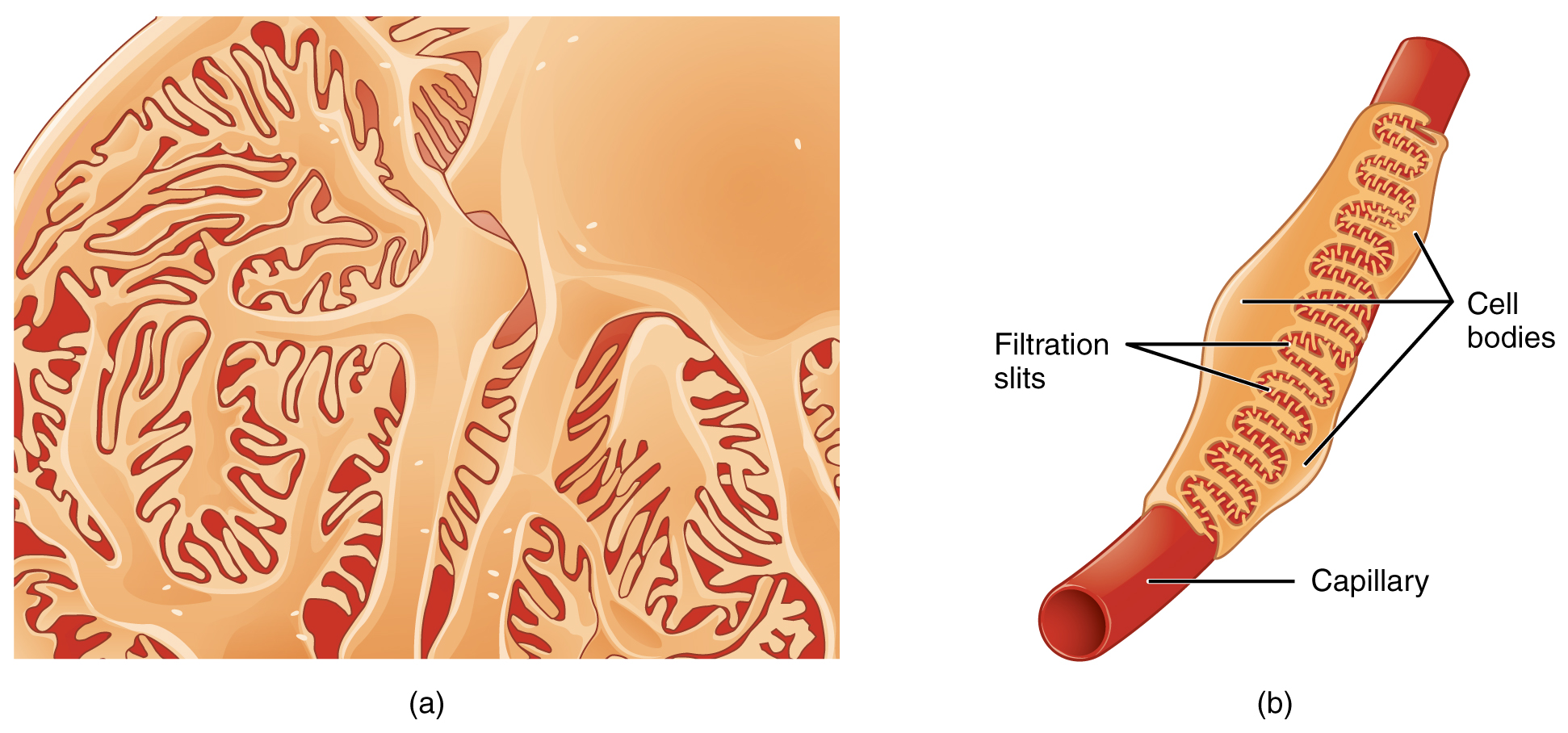
Microscopic Anatomy of the Kidney · Anatomy and Physiology

References in Podocyte Biology for the Bedside American Journal of

Nck Links Nephrin to Actin in Kidney Podocytes Cell

Podocyte définition et explications

Podocyte foot process architecture and proteins involved in hereditary

Podocytes structure and function YouTube

What Are Podocytes Astral Projection
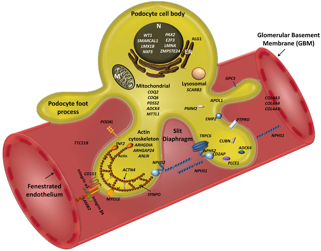
Frontiers Genes and Podocytes New Insights into Mechanisms of

A model of podocyte maintenance and re generation. Parietal epithelial
Related Post: